VetMED Emergency & Specialty Veterinary Hospital

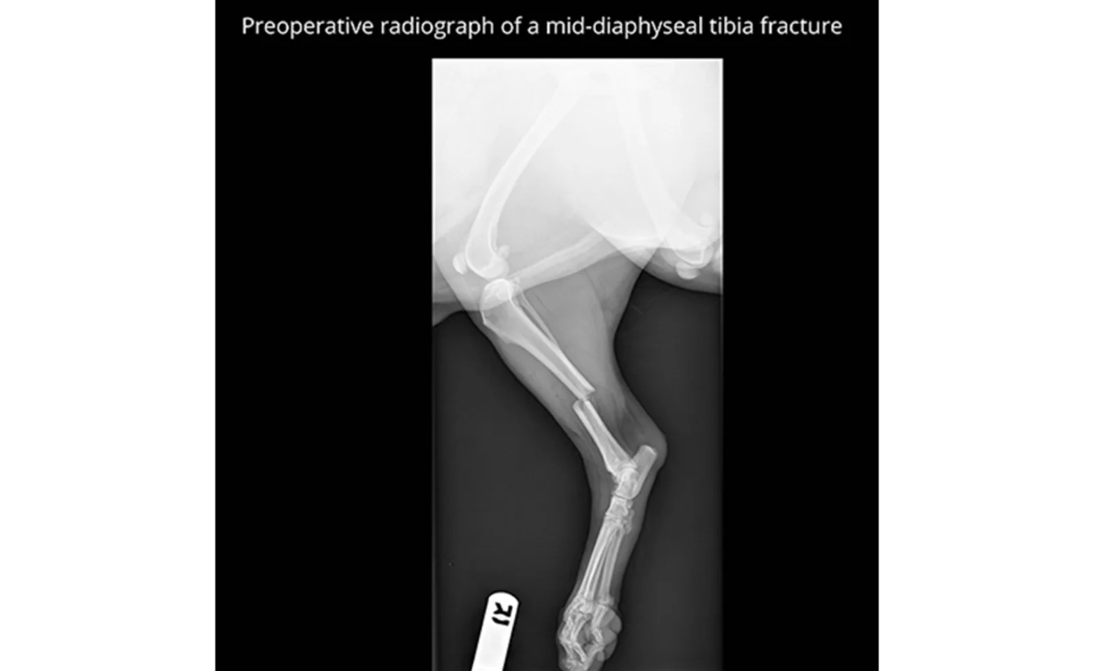
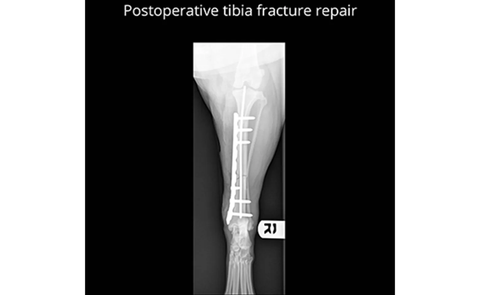
A fracture can seem like a devastating injury for your pet. Fortunately, the majority of fractures are fixable and carry a good long-term prognosis. The method of fracture repair will depend on the bone or bones that are involved. The most common methods of fracture stabilization are performed with bone plates, screws, and pins. The average time for a fracture to heal is about 8-10 weeks but may be shorter for young patients, or slightly longer for geriatric patients. More recently, certain fractures are being repaired in a minimally invasive fashion. This method uses intra-operative imaging, called fluoroscopy, to evaluate the bone while it is being aligned and stabilized with implants. With the use of fluoroscopy, incisions can be much smaller and the blood supply to the bone is better preserved. This type of fracture repair is usually referred to as “Minimally Invasive Plate Osteosynthesis” or “MIPO”.

A fracture and patient must meet very specific criteria to be a candidate for non-surgical management with a cast or splint. A very common fracture seen in veterinary patients is a break in the lower forelimb bones (radius and ulna), and usually this occurs in small breed dogs. This is a fracture that should always be managed surgically, as studies have shown a very high rate of complications when they are managed non-surgically.
It is recommended that fractures be repaired by those with knowledge of bone biology, healing, and fracture biomechanics, and our board-certified surgeons at VetMED have a great deal of experience and interest in this field.
For more information about fracture repair services at VetMED or if you would like to speak with one of our board-certified surgeons, please call (602) 697-4694 to make an appointment.

